S'il vous plaît
actualisez votre navigateur
pour avoir l'opportunité d'utiliser pleinement Internet, des millions de personnes l'ont déjà fait il y a longtemps.

Summary of the article
Qu'est-ce qu'une échographie/un doppler ?
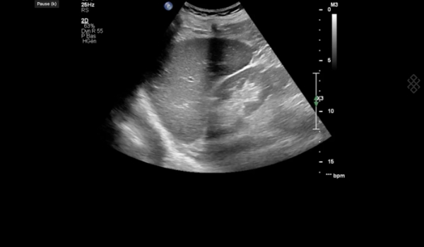
L'échographie permet d'explorer différents organes du corps en plaçant une sonde sur la peau. Cette sonde émet des ondes ultrasonores qui sont réfléchies par les tissus explorés puis détectées sous forme d'échos par cette même sonde et traduites en images interprétables par un ordinateur.
Elle est utilisée pour rechercher des anomalies sur les organes de l'abdomen (le foie, la vésicule biliaire, la rate, le pancréas et les reins), les organes génitaux (prostate, testicules, utérus, ovaires) et le cœur. Elle permet également l'exploration des tissus mous, des ligaments, des tendons et des muscles.
Technique indispensable au cours d'une grossesse, elle est l'examen de référence pour suivre le développement du fœtus et dépister d'éventuelles anomalies.
Le doppler permet, avec la même technique, l'exploration du sang circulant dans les veines et les artères. Indiqué en cas de suspicion de phlébite (caillot bloqué dans une veine) ou de plaques d'athérome (rétrécissement du calibre d'une artère), il est un bon moyen d'étudier les perturbations du flux sanguin et d'évaluer la vascularisation des organes.
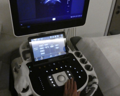
L'appareil utilisé pour réaliser une échographie ou un doppler s'appelle un échographe. Il est constitué d'une sonde, d'un panneau de commande, d'un écran vidéo et d'un système informatique.
Comment se déroule une échographie/un doppler ?
L’échographie est faite par un médecin radiologue ou autre spécialiste. Les sages-femmes sont aussi en mesure d'effectuer cet examen lorsqu'il s'agit de suivre l'évolution d'une grossesse.
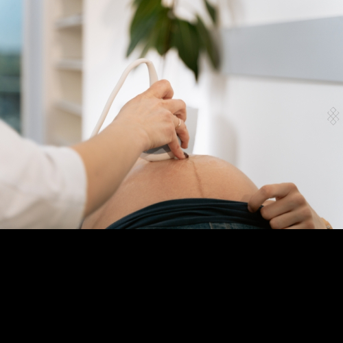
Suivant la région à examiner, vous serez préparé(e) différement. Le médecin vous précisera quels vêtements enlever afin d'avoir accès à votre peau pour y poser la sonde, en cas d'échographie transcutanée. Dans certains cas, l'échographie peut se faire par voie endovaginale ou endorectale.
Pendant l'examen, vous serez allongé(e) le plus souvent sur le dos. Du gel d'échographie sera appliqué sur la sonde et sur votre peau pour améliorer la transmission des ultrasons et le médecin vous commentera en direct ce qu'il observe.
L'échographie/doppler dure en moyenne entre 10 et 30 minutes selon la région à examiner. Ensuite vous repartirez avec un compte-rendu que vous transmettrez à votre médecin prescripteur.
Se préparer à une échographie/un doppler
Toutes les modalités de préparation (être à jeun, avoir la vessie pleine...) vous seront expliquées lors de votre prise de rendez-vous. Bien souvent, elles sont écrites sur votre convocation qui vous a été remise par la secrétaire médicale ou par voie dématérialisée. Il faudra alors bien respecter ces consignes. Votre examen pourrait ne pas être réalisé si vous n'avez pas fait une bonne préparation, les conditions ne pouvant garantir une analyse optimale et donc des résultats fiables.
Le jour de l'examen, pensez à présenter au secrétariat les documents suivants :
- Votre carte vitale et mutuelle
- La demande de votre médecin (ordonnance, compte-rendu d'examen)
- Vos anciens comptes-rendus et les images en rapport avec l'affection qui permettront une comparaison et un meilleur suivi (radiographie, échographie, scanner, IRM...)
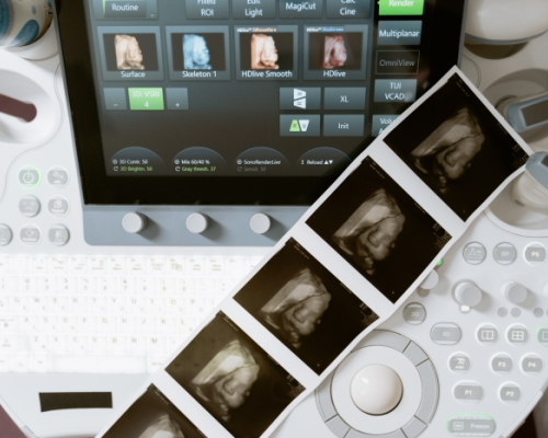
Interprétation des images
L'interprétation des images ainsi que leur enregistrement se fait en même temps que l'échographie. Le médecin vous montre et vous explique les images sur l'écran.
Pensez à l'informer de tout antécédent médical pouvant l'aider au diagnostic et n’hésitez pas à poser toutes vos questions.
Dans le cas où, les résultats de l'échographie ne sont pas suffisants, des examens complémentaires pourront être prescrits par le radiologue.
Quels sont les risques d'une échographie ?
Les ondes ultrasonores sont sans danger.
Il n'y a donc pas de contre-indication ni aucune complication liée à la réalisation d’une échographie simple qu'elle soit transcutanée, endovaginale ou endorectale.
Par ailleurs, les échographies couplées à une endoscopie peuvent présenter quelques risques. Ils sont liés notamment à l'anesthésie et aux infections.