
Qu'est-ce qu'une mammographie ?
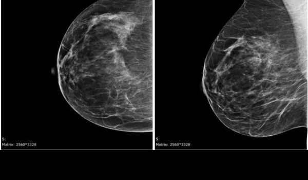
La mammographie permet de radiographier les seins à l'aide d'un tube radiogène. Le faisceau de rayons-X émis du tube traverse le tissu mammaire et donne une image radiologique contrastée.
Grâce à elle, des anomalies des tissus telles que des opacités, des micro-calcifications ou des tumeurs sont détectées.
Elle intervient en premier recours pour le diagnostic du cancer du sein notamment avec le dépistage organisé prévu tous les 2 ans entre 50 et 74 ans.
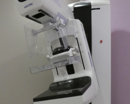
La salle de mammographie comprend un appareil appelé mammographe. Il est constitué d'un capteur plan, sur lequel sera posé le sein, ainsi que d'un tube à rayons-X, le tout articulé selon plusieurs axes. Le pupitre de commande est séparé par un paravent plombé pour protéger le manipulateur ou la manipulatrice en radiologie.
Comment se déroule une mammographie ?
La mammographie sera programmée de préférence durant les dix premiers jours du cycle. C'est dans cette première partie du cycle menstruel que les seins sont moins sensibles.
Vous serez amené(e) à enlever seulement le haut de votre tenue. Pensez donc à porter des vêtements adaptés. Évitez de mettre le jour de l'examen, des produits de type crème, poudre, parfum ou déodorant sous les aisselles ou sur les seins. Ceux-ci peuvent parfois gêner la réalisation de l'examen ou l'interprétation des résultats.
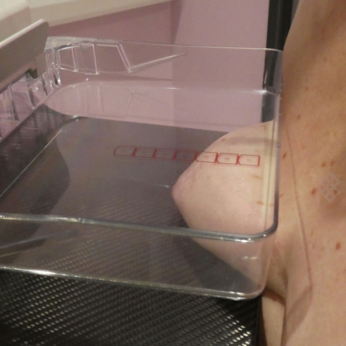
Pour réaliser les clichés (de face, en oblique et +/- de profil) le sein est placé sur le mammographe puis est progressivement comprimé. Pour éviter tout mouvement, vous devrez couper votre respiration le temps du cliché.
Il faut compter en moyenne 20 minutes pour une prise en charge en mammographie.
A l'issue de la mammographie, le médecin radiologue vous examine. Il vérifie l’aspect de vos seins et recherche par la palpation une éventuelle tuméfaction ou des ganglions anormaux.
En cas de besoin, la mammographie peut être complétée par une échographie mammaire qui sera faite dans la foulée.
Se préparer à une mammographie
Aucune préparation n'est nécessaire. Pas besoin d'être à jeun, vous pouvez donc manger, boire et prendre vos médicaments comme d’habitude.
Le jour de l'examen, pensez à présenter au secrétariat les documents suivants :
- Votre carte vitale et mutuelle
- La demande de votre médecin (ordonnance, compte-rendu d'examen)
- Vos anciennes mammographies, échographies mammaires (et tout autre résultat qui pourrait être utile au radiologue) qui permettront une comparaison et donc un meilleur suivi.
Interprétation des images
Un commentaire vous sera fait à la fin de l'examen et les résultats vous seront remis s'il ne s'agit pas d'un dépistage organisé qui nécessite une deuxième lecture. Celle-ci, effectuée par un autre radiologue, sert à confirmer les résultats de la mammographie. Dans ce cas, les clichés seront envoyés à l'organisme de dépistage qui, après la deuxième lecture, vous enverra vos clichés et le compte-rendu.
Si nécessaire et au vu des résultats, il vous sera prescrit des examens supplémentaires, pour compléter votre bilan (échographie, IRM mammaire, ponction mammaire ou biopsie percutanée).
Quels sont les risques d'une mammographie ?
La mammographie utilise les rayons-X, c'est une technique d'imagerie irradiante. Cependant les faibles doses reçues lors d'une mammographie sont largement acceptables et rendent le risque faible par rapport au bénéfice médical.
La contre-indication majeure est la grossesse. Il est donc impératif de signaler toute grossesse éventuelle ou en cours pour que les précautions soient prises.