
Qu'est-ce qu'une endoscopie ?
L'Endoscopie permet de visualiser l'intérieur d'un organe creux. Pratiquée par un médecin spécialiste, elle permet d'accéder à un organe pour son analyse visuelle. Introduit à l'intérieur de l'organisme par les voies naturelles ou à l'aide de petites incisions, l'endoscope retransmet les images sur un écran.
On distingue deux catégories d'endoscopie, celle à visée diagnostique à la recherche des causes d'un trouble, et celle à visée thérapeutique dans l'objectif de traiter une pathologie. Les différentes dénominations de l'endoscopie en fonction des organes explorés :
- L'arthroscopie pour l'exploration des articulations
- La gastroscopie ou la fibroscopie digestive haute pour le système digestif
- La cœlioscopie pour l'exploration de l'abdomen
- La bronchoscopie pour l'exploration des bronches
- La coloscopie pour l'exploration du côlon
- La cystoscopie pour l'exploration de la vessie
- L'hystéroscopie pour l'exploration de l'utérus
L'endoscopie n'utilise pas de rayon-X, ce n'est donc pas une technique irradiante.
L'appareil utilisé est appelé endoscope ou fibroscope. Il est constitué d'un tube rigide ou souple, au bout duquel sont fixées une lampe et une caméra. Pour chaque organe, il existe un endoscope adapté.
Comment se déroule une endoscopie ?
Il existe différentes catégories d'endoscopie qui nécessitent chacune une prise en charge particulière.
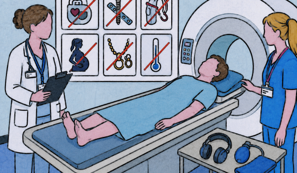
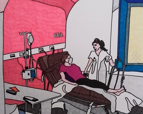
Généralement, vous êtes pris en charge par le personnel médical qui vous demande d'ôter vos vêtements, vous installe sur la table d'examen puis vous met une perfusion.
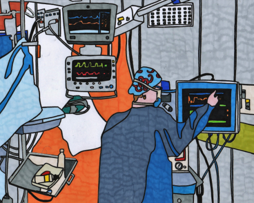
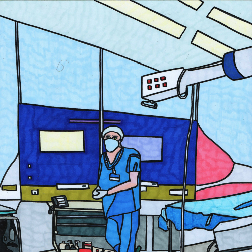
Selon l'organe étudié et le type d'examen prescrit, une anesthésie locale ou générale peut être réalisée. L'examen se déroule dans les conditions optimales de sécurité et d'hygiène.
Si besoin, pour faciliter le déplacement du dispositif médical, de l'air pourra être insufflé ce qui permettra de décoller les parois. D'autres accessoires peuvent être également utilisés pour permettre de prélever, de sectionner ou de traiter les tissus.
L'examen dure de 30 minutes à une heure environ. Après l'endoscopie, une surveillance de votre état de santé peut s'avérer nécessaire, cela implique une hospitalisation allant d'un demi journée à 48 heures.
Se préparer à une endoscopie
Lors de la prise de rendez-vous, pensez à mentionner vos traitements, surtout si vous êtes sous anti-coagulants, ainsi que vos antécédents médicaux.
Si l'intervention est prévue sous anesthésie vous devrez au préalable consulter un médecin anesthésiste, à peut prés 10 jours avant l'examen. D'autres bilans peuvent vous être prescrits (bilan sanguin, électrocardiogramme, radio pulmonaire...).
En cas d’anxiété non soulagée par les explications et les conseils des soignants, le médecin pourra décider de vous administrer un tranquillisant.
La plupart du temps il faut être à jeun quatre à six heures avant l'examen, vous ne devez donc ni manger, ni boire ni fumer.
Le jour de l'examen, pensez à présenter au secrétariat les documents suivants :
- Votre carte vitale et mutuelle
- La demande de votre médecin (ordonnance, compte-rendu d'examen)
- Vos anciens comptes-rendus et les images en rapport avec l'affection qui permettront une comparaison et un meilleur suivi (radiographie, échographie, scanner, IRM...)
- Vos résultats d'analyses de sang

Interprétation des images
Les résultats vous seront donnés à la fin de l'examen par le médecin spécialiste. Il vous fera un commentaire, vous expliquera la conduite à tenir pour la suite et rédigera un compte-rendu écrit pour vous et votre médecin traitant.
Quels sont les risques d'une endoscopie ?
Les complications d’un examen endoscopique restent exceptionnelles, parmi celles-ci, les infections sont très rares.Cependant, si des symptômes apparaissent (douleur, fièvre, perte de sang...) il faudra le signaler aux soignants, si vous êtes hospitalisé.e, ou auprès de votre médecin traitant.
L'anesthésie générale constitue un risque potentiel. La consultation pré-anesthésique permettra de la réaliser dans les conditions optimales.