
S'il vous plaît
actualisez votre navigateur
pour avoir l'opportunité d'utiliser pleinement Internet, des millions de personnes l'ont déjà fait il y a longtemps.

La radiologie interventionnelle
La radiologie interventionnelle comprend l'ensemble des techniques d'imagerie permettant le traitement ou le diagnostic invasif de différentes maladies. Elle utilise les rayons-X, les ultrasons ou la résonance magnétique.
Summary of the article
Qu'est-ce qu'un examen interventionnel ?
La radiologie interventionnelle permet d'accéder et de visualiser différents organes afin d'effectuer un acte diagnostique ou thérapeutique. Réalisée sous guidage radiologique (radioscopie ou fluoroscopie) et sous anesthésie locale ou générale, elle est une bonne alternative à la chirurgie bien plus invasive.
Son domaine d'intervention est large et couvre un ensemble de spécialités. Ainsi les médecins radiologues pratiquent la radiologie interventionnelle alors que les cardiologues interviennent dans la cardiologie interventionnelle et les neurologues dans la neurologie interventionnelle. Elle touche d'autre domaines médicaux comme l'urologie, le vasculaire, la gynécologie et sénologie, l'ostéoarticulaire, la gastro-entérologie ou encore la pédiatrie.
Les différentes techniques interventionnelles peuvent être couplées aux techniques chirurgicales et endoscopiques ce qui permet, en même temps que de progresser vers les organes sous guidage radioscopique, d'y accéder directement par la chirurgie ou l'endoscopie.
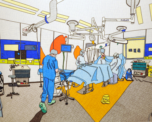
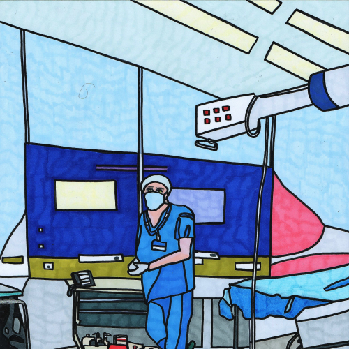
La salle de radiologie interventionnelle est constituée d'un tube à rayons-X qui s'articule autour du patient pour retranscrire en direct les images par l'acquisition de micrographie ou scopie. Elle est également munie d'une table d'examen ainsi que d'un équipement spécial stérile pour les actes invasifs.
Lorsque les actes interventionnels sont réalisés dans une salle d'IRM ou de scanner, les salles sont spécialement équipées pour respecter le cadre médical strict.
Comment se déroule un examen interventionnel ?
Il existe plusieurs actes interventionnels qui nécessitent chacun une prise en charge particulière.
Généralement, vous êtes pris en charge par le personnel médical qui vous demande d'ôter vos vêtements, vous installe sur la table d'examen puis vous met une perfusion.
Selon l'organe étudié et le type d'examen prescrit, une anesthésie locale ou générale peut être réalisée. L'examen se déroule dans les conditions optimales de sécurité et d'hygiène.
L'examen dure de 30 minutes à une heure environ. Après, une surveillance de votre état de santé peut s'avérer nécessaire, cela implique une hospitalisation allant d'un demi journée à 48 heures.
Pendant l'examen, un produit de contraste est injecté et des clichés sont réalisés pour mettre en évidence des pathologies au niveau des vaisseaux, d'une articulation, ou autre organe étant la cible de l'intervention. Un acte thérapeutique pourra être couplé en fonction des images obtenues et des conditions favorables ou non.
N'hésitez pas à signaler au personnel médical tout problème pendant l'examen.
Se préparer à un examen interventionnel
Lors de la prise de rendez-vous, pensez à mentionner vos traitements, surtout si vous êtes sous anti-coagulants, ainsi que vos antécédents médicaux. Un bilan sanguin vous sera prescrit.
Si vous avez des allergies, le médecin vous prescrira des comprimés à prendre la veille et le jour de l'examen.
Si l'intervention est prévue sous anesthésie vous devrez au préalable consulter un médecin anesthésiste, à peut prés 10 jours avant l'examen. D'autres bilans peuvent vous être prescrits (bilan sanguin, électrocardiogramme, radio pulmonaire...).
La plupart du temps il faut être à jeun quatre à six heures avant l'examen, vous ne devez donc ni manger, ni boire ni fumer.
Le jour de l'examen, pensez à présenter au secrétariat les documents suivants:
- Votre carte vitale et mutuelle
- La demande de votre médecin (ordonnance, compte-rendu d'examen)
- Le produit de contraste s'il vous a été prescrit
- Vos anciens comptes-rendus et les images en rapport avec l'affection qui permettront une comparaison et un meilleur suivi (radiographie, échographie, scanner, IRM...)
- Vos résultats d'analyses de sang
Quel produit de contraste ?
Le produit utilisé lors d'un examen interventionnel dépend de la technique d'imagerie.
Lorsque les appareils utilisent les rayons-X pour visualiser les organes, le produit de contraste injecté est à base d'iode. Dans le cas de l'imagerie par résonance magnétique, une solution à base de gadolinium sera utilisé.
L'injection est un acte très courant et il est généralement très bien toléré.
La plupart du temps, il vous sera prescrit un dosage de créatinine pour vérifier votre fonction rénale.
Lors de l'injection du produit iodé, il se peut que vous ressentiez une sensation de chaleur dans tout le corps avec plus ou moins un goût étrange dans la bouche. Cela durera en moyenne moins d’une minute.
Généralement bien supportée, il existe quelques faibles risques liés à l'injection :
- L'hématome provoqué par la pose d'une voix veineuse périphérique. Sans gravité, il se résorbera en quelques jours
- La fuite sous-cutanée du produit de contraste. Elle est due à la pression lors de l'injection et ne présente généralement pas de suite grave.
- Des nausées voir des vomissements.
- De l'urticaire de manière plus rare.
- Une réaction allergique rare de type eczéma, asthme ou troubles cardio-respiratoires
- Une Fibrose Systémique Néphrogénique qui intervient exceptionnellement en cas d'insuffisance rénale.
Ces réactions imprévisibles d'intolérance au produit de contraste arrivent, plus fréquemment, chez les patients ayant des antécédents allergiques. Elles sont généralement transitoires et sans gravité.
Selon la gravité de la réaction, il vous sera administré un traitement et vous serez orienté(e) pour un dépistage allergologique.
Dans tous les cas (sauf avis médical contraire), s'il vous a été administré un produit de contraste, buvez deux litres d’eau après l'examen. Cela aidera à éliminer rapidement le médicament.

Interprétation des images
Les résultats vous seront donnés à la fin de l'examen par le médecin spécialiste. Il vous fera un commentaire, vous expliquera la conduite à tenir pour la suite et rédigera un compte-rendu écrit pour vous et votre médecin traitant.
Si un service de télé-radiologie est mis en place, les résultats vous seront communiqués via une plateforme web. Vous disposerez alors d'identifiants pour consulter vos compte-rendu et images en ligne à l'aide d'une connexion sécurisée à un portail patient.