S'il vous plaît
actualisez votre navigateur
pour avoir l'opportunité d'utiliser pleinement Internet, des millions de personnes l'ont déjà fait il y a longtemps.

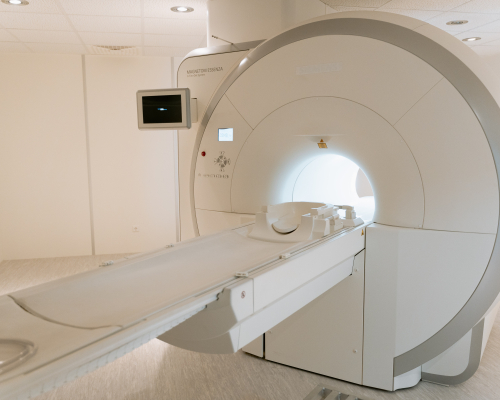
L’imagerie par résonance magnétique
L’imagerie par résonance magnétique (IRM) est une technique d’imagerie médicale qui utilise un champ électromagnétique. Elle permet une meilleure exploration des organes que l'échographie, les radiographies standards ou le scanner. La reconstruction des images se fait dans les trois plans de l'espace.
Summary of the article
Qu'est-ce qu'une IRM ?
L'IRM permet de visualiser de manière très précise presque toutes les parties du corps en utilisant les protons présents dans les molécules d'eau. Les atomes d'hydrogène réagissent aux ondes de radiofréquence qui sont envoyées par la machine en absorbant l'énergie. Excités, ils restituent ensuite l'énergie accumulée et produisent un signal qui est lu et transformé en une image anatomique.
Hormis les poumons qui ne sont pas explorés par cette technique, l'IRM est d'une grande efficacité pour analyser le cerveau, la colonne vertébrale, les articulations et les tissus mous. Ainsi elle permet de déceler des lésions infectieuses ou inflammatoires, des anomalies des vaisseaux, des lésions ligamentaires ou méniscales et des tumeurs.
L'IRM n'utilise pas de radiations ionisantes comme en radiologie ou au scanner, elle n'est donc pas irradiante.
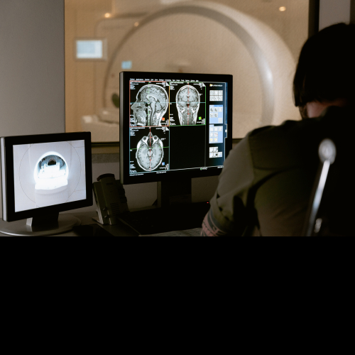
Elle est composée d'un tunnel qui comprend à l'intérieur un gros aimant ainsi que différentes antennes qui serviront à explorer une région du corps. En dehors de la salle d'examen, une console informatique permet aux professionnels médicaux et paramédicaux de programmer les séquences d'images ainsi que leur interprétation.
Comment se déroule une IRM ?
En fonction de l'organe à étudier, comptez 15 à 45 minutes pour passer une IRM. Sous la responsabilité du médecin radiologue, vous serez pris en charge par un manipulateur ou une manipulatrice en radiologie.
Sauf indication contraire, pensez à aller aux toilettes pour éviter toute envie pendant l'examen qui peut être un peu long.
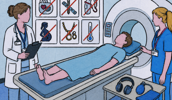
Avant d'entrer dans la salle, vous serez interrogé(e) sur vos antécédents médicaux. Le professionnel de santé s'assure que vous ne présentez pas de contre-indication absolue ou majeure. Il est important de bien signaler :
- Si vous avez un stimulateur cardiaque ou tout autre système électronique implantable
- Si vous avez été opéré du cœur ou de la tête
- Si vous avez un corps étranger métallique dans le corps (implant oculaire, prothèse, clips chirurgicaux, éclat de métal dans les yeux…)
- Si vous souffrez d'une insuffisance rénale, de diabète
- Si vous avez déjà fait une allergie à un produit de contraste à base de gadolinium
- Une grossesse éventuelle ou en cours
- Si vous êtes claustrophobe (peur d'être confiné(e), enfermé(e))
Se préparer à une IRM
A l'exception de quelques examens, aucune préparation n'est nécessaire. Pas besoin d'être à jeun, vous pouvez donc manger, boire et prendre vos médicaments comme d’habitude.
Le jour de l'examen, pensez à présenter au secrétariat les documents suivants :
- Votre carte vitale et mutuelle
- La demande de votre médecin (ordonnance, compte-rendu d'examen)
- Le produit de contraste qui vous a été prescrit en cas d'examen injecté
- Vos anciens comptes-rendus et les images en rapport avec l'affection qui permettront une comparaison et un meilleur suivi (radiographie, échographie, scanner, IRM...)
- Votre carte de dispositif médical implantable si vous avez été opéré(e)
- Vos résultats d'analyses de sang
Portez de préférence des vêtements n'ayant pas de fermeture éclair ou des boutons pressions, évitez de mettre des bijoux et des épingles à cheveux, sinon vous devrez les enlever.
Produit de contraste à l'IRM
Certaines indications nécessitent l'injection d'un produit de contraste. Le produit utilisé à l'IRM est à base de Gadolinuim, il permet de rendre visibles les éléments pathogènes sur les images.
L'injection est un acte très courant à l'IRM qui est généralement très bien toléré. Des réactions imprévisibles d'intolérance au produit de contraste sont très rares et arrivent plus fréquemment chez les patients ayant des antécédents allergiques. Elles sont généralement transitoires et sans gravité.
La plupart du temps, il vous sera prescrit un dosage de créatinine pour vérifier votre fonction rénale. Si vous souffrez d'insuffisance rénale, l'injection sera (sauf nécessité absolue) annulée.
Généralement bien supportée, il existe quelques faibles risques liés à l'injection :
- L'hématome provoqué par la pose d'une voix veineuse périphérique. Sans gravité, il se résorbera en quelques jours
- La fuite sous-cutanée du produit de contraste. Elle est due à la pression lors de l'injection et ne présente généralement pas de suite grave
- Des nausées voir des vomissements.
- De l'urticaire de manière plus rare
- Une réaction allergique rare de type eczéma, asthme ou troubles cardio-respiratoires
- Une Fibrose Systémique Néphrogénique qui intervient exceptionnellement en cas d'insuffisance rénale
Selon la gravité de la réaction, il vous sera administré un traitement et vous serez orienté(e) pour un dépistage allergologique.
Dans tous les cas (sauf avis médical contraire), s'il vous a été administré un produit de contraste, buvez deux litres d’eau après l'examen. Cela aidera à éliminer rapidement le médicament.

Interprétation des images
L'interprétation des images peut se faire à la fin de l'examen si un radiologue est en mesure de les analyser immédiatement ou plus tard lorsqu'un service de télé-radiologie est mis en place.
Avant de rentrer chez vous, le secrétariat vous remettra le compte-rendu et les images souvent gravés sur un CD-ROM. Si l'impression des résultats sous cette forme n'est pas possible ou si le centre a opté pour la télé-radiologie, vous disposerez alors d'identifiants pour consulter vos résultats en ligne à l'aide d'une connexion sécurisée à un compte patient.
Quels sont les risques d'une IRM ?
Il n'y a pas de risque biologique connu lié au champ magnétique.
Les risques liés à l'injection de produit de contraste sont faibles : Voir Produit de contraste à l'IRM.
Le port d'une pile cardiaque, valve cardiaque ou tout corps étranger métallique implanté dans votre organisme (notamment dans la tête) représente un facteur de risque majeur s'il n'est pas signalé à l'équipe médicale.